[PUBLICATION] Multicenter study of organ-at-risk protection tools in CT scanning: radiation protection and image quality

Presented at JFR, DGMP and RSNA, discover the summary of this work below.

Introduction :

CT scans accounted for 12.8% of all imaging examinations conducted in 2017. However, these scans represented more than 70% of the collective effective dose. To reduce patient exposure, manufacturers have implemented several dose optimization tools. Among these techniques, we can mention organ-based tube current modulation (OBTCM) as ODM, OEM and X-CARE, which supposed to reduce the dose on the anterior face of the patients. Nevertheless, the only available dose indicators (CTDI, DLP) do not allow to study the functioning of these tools. Thus, the objective of this study was to assess the relevance of the organ dose protection tools proposed by each multi-slice CT scanner manufacturer, by estimating their impact in terms of dosimetry and image quality, specifically for head examinations.

Material & Method :

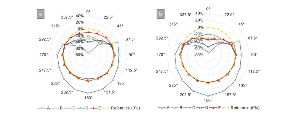
Five CT scanners installed in four radiology services were evaluated: Siemens Somatom Go Top, GE (Revolution CT and Revolution Maxima), Canon (Aquilion Genesis, and Aquilion Prime). Measurements were performed using two head protocols on a HEAD16 phantom (16 cm diameter, PMMA): a “standard” protocol to compare the devices with each other and a “clinical” protocol used in routine practice. To quantify the dose reduction obtained using the OBTCM tools, the scintillating fiber detector IVInomad, was placed on the inner part of the gantry at 16 measurement positions, placed all around the gantry (see Figure 1). Image quality was evaluated using standard metrics (SD, SNR, NPS) on the HEAD16 phantom images on four regions of interest, positioned in pairs respectively on the anterior and posterior parts of the phantom. Statistical analysis was performed (Wilcoxon test, Bland-Altman) on the measurements obtained with and without the OBTCM tools to evaluate the significance of image quality differences.

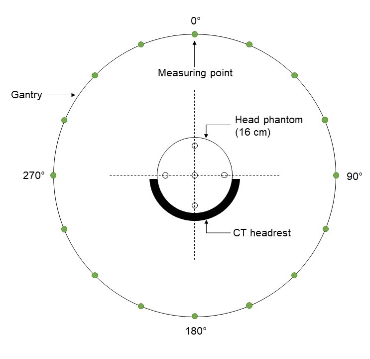
Figure 1: Schema of the dose measurement protocol. Dots represent the 16 measuring points using the IVInomad detector.

Results:

As it is shown in Figure 2, the dose on the anterior face of the patient is reduced by 10.9% to 73.8% depending on the protocol and scanner used. The dose on the posterior face remains unchanged on GE and Canon scanners. However, on Siemens CT scanner, the dose on the posterior face increases by approximately 39%. The noise (SD) increases by 2.4% to 10.4% depending on the scanner and protocol studied. In absolute terms, this corresponds to a maximum increase of 0.5 HU. Similarly, the SNR decreases between 2.6% and 9.6%, resulting in an absolute maximum decrease of 2.5 HU. These differences are considered statistically significant (p-value < 0.05). The analysis of the NPS showed that the noise texture was not affected.

Figure 2: Dose reduction rates measured in the five CT scanners using: the standard protocol (a) and the routine protocol (b). The dotted line represents a null difference between doses measured with and without OBTCM. (A): Siemens Somatom Go Top. (B): GE Revolution CT. (C): GE Revolution Maxima. (D): Canon Aquilion Genesis. (E): Canon Aquilion

Conclusion:

The OBTCM tools significantly reduce the dose on the patient’s anterior face. In the case of this specific study on head examinations, these tools reduce the exposure to the patient’s anterior face by up to 73.8%, depending on the CT scanner and the protocol used. However, an impact on image quality is observed, with a maximum increase of 0.5 HU in standard deviation and a maximum decrease of 2.5 HU in SNR. maximum. . .

Share this article

Share on Facebook
Share on Linkdin

Let's talk together!

Are you a health care professional?

Stay informed with our free relationship program for outreach professionals.

Want to become a partner?

Distribution, R&D, clinical tests…
Contact us!