[PUBLICATION] "Evaluation of a Scintillating Plastic Optical Fiber Device for Measuring kV-Cone Beam Computed Tomography Dose"
Background: Justification of imaging procedures such as cone beam computed tomography (CBCT) in radiotherapy makes no doubt. However, the CBCT composite dose is rarely reported or optimized, even though the repeated CBCT cumulative dose can be up to 3% of the prescription dose This study aimed to evaluate the performance and utility of a new plastic scintillating optical fiber dosimeter for CBCT dosimetric quality assurance (QA) applications before a potential application in patient composite CBCT dosimetry.
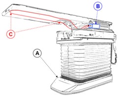
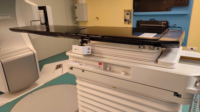
Figures: Left: Schematic depiction of the components of the measurement device and its setup on the treatment unit (A) the foot of the couch of the accelerator. (B) The signal treatment unit composed of a battery and bluetooth emitter. (C) The optical fiber, which is securely beneath the couch top. The two arrows point to the beginning and the end of the sensing part of the probe; Right: IVIcbct system installed on a Varian TrueBeam unit at the Radiotherapy department of CHR Metz-Thionville.
The fiber impact on the fluence and dose delivered was respectively assessed with an electronic portal imaging device (EPID) and EBT3 Gafchromic® film. The presence of artifacts was visually evaluated on kV images. The dosimeter performances were determined for various acquisition parameters by comparison with ionization chamber values.
Results: The maximum impact of the fiber on the fluence measured by the EPID was −1.2% for the 6 MV flattening filter-free beam. However, the fiber did not alter the film dose profile when measured for all the beams tested. The fiber was not visible at energies ≥80 kV and was merely visible on the CBCT images. When the rate of images per second or mA was changed, the maximum relative difference between the device and the ionization chamber CTDIs was<5%. Changing collimation led to a −7.2% maximum relative difference with an absolute dose difference that was insignificant (−0.3 mGy). Changing kV was associated with a −8.7% maximum relative difference, as published in the literature.
Conclusions: The dosimeter may be a promising device for CBCT recurrent dosimetry quality control or dose optimization. What’s more, given the sensor’s length (1m), CBDI measurements are much easier and quicker than those made with a standard pencil ion chamber. According to these results, further developments are in progress in order to adapt the solution to the measurement of patient composite CBCT doses.
You can find our previous articles on the validation of the system for the scanner modality and on its complete characterization here:
https://www.mdpi.com/1424-8220/23/5/2614
https://www.mdpi.com/1424-8220/22/1/90