
[PUBLICATION] The impact of acceleration factors in 4D flow MRI
Original title: “Accelerated sequences of 4D flow MRI using GRAPPA and compressed sensing:
A comparison against conventional MRI and computational fluid dynamics” published online in
August 2022 in the journal Magnetic Resonance in Medicine ▸https://doi.org/10.1002/mrm.29404
Interest of the study
So-called “4D flow MRI” sequences provide an image of the
blood flow velocity, both within a given volume and over the course of the cycle
cardiac [1]. This promising technique provides non-invasive access to
hemodynamic biomarkers such as flow rate, maximum velocities or even
derived parameters (e.g. parietal friction, turbulence kinetic energy). However,
the clinical use of 4D flow MRI remains underdeveloped due to the long duration of its use.
acquisitions. Various techniques have been developed to speed up acquisition time:
parallel imaging (e.g. GRAPPA, SENSE) or, more recently, Compressed Sensing [2,3].
The main objective of this study is to investigate the measurement errors induced by the
GRAPPA and Compressed Sensing accelerations for flow characterization
obtained with 4D flow MRI, in a well-controlled environment featuring
complex flow patterns typical of those seen in the cardiovascular system.
Method
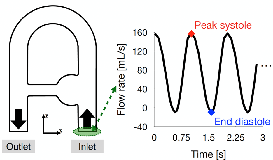
- Test object (phantom) with rigid wall [4], receiving a pulsed flow from a programmable pump (see diagram opposite).
- Acquisitions on a 1.5 Tesla MRI scanner (MAGNETOM Sola, Siemens Healthcare, Erlangen, Germany)
- 1 fully sampled (FS) acquisition - duration 43 minutes
- 3 GRAPPA acquisitions, accelerated by a factor of 2, 3 and 4 - duration 21, 15 and 11 minutes, respectively
- 1 Compressed Sensing (CS) acquisition, accelerated by a factor of 7.6 - duration 6 min
- Reconstruction of 20 cardiac phases (25 for CS)
- Computational Fluid Dynamics (CFD) simulations [5], with the flow rate obtained from the various 4D flow MRI acquisitions imposed as input
The MRI acquisitions and MFN simulations were then post-processed to allow their
comparison following the recommendations of Puiseux et al. 2019 [4]. To compare
The following quantities have been evaluated:
- Comparison of voxel-to-voxel velocity (voxel = 3D spatial point of the MRI image)
- Speed profiles
- Flow rates
- Maximum speeds
Results
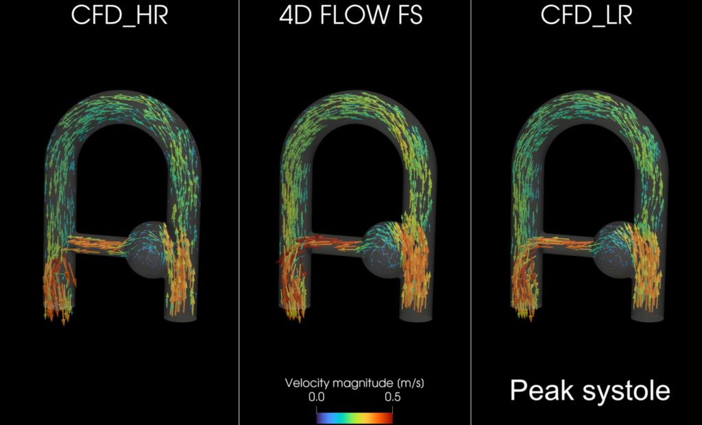
Qualitatively, similar hemodynamic patterns were observed for all the
modalities. In the video opposite, the non-accelerated acquisition (4D FLOW FS) is compared with the 4D FLOW FS.
MFN simulation (CFD_HR), and to the same simulation under-resolved to match the
MRI voxels (CFD_LR).
Quantitatively, it was found that :
- The voxels close to the ghost walls are the ones with the most differences between the modalities.
- Compared to MFN, speed profiles and maximum speeds tend to be higher in areas associated with high speeds and/or acceleration.
- The correction of eddy currents in MRI images appears essential to guarantee consistent throughputs in terms of mass conservation.
Conclusion
This study shows that highly accelerated Compressed Sensing acquisition enables
obtain in vitro results very similar to those obtained with a non-accelerated sequence
or with conventional GRAPPA-type accelerations (factors 2 or 3). However, it
artifacts are inherent to the 4D flow MRI acquisition procedure: artifacts from
spatial and velocity displacement, intravoxel phase shifting, partial volume effects… Although
also has its limitations, the MFN appears to be a tool that can be used to study
discrepancies.
[1] Markl M, Frydrychowicz A, Kozerke S, Hope M, Wieben O. 4D flow MRI. J Magn Reson
Imaging. 2012;36(5):1015-1036. doi:10.1002/jmri.23632
[2] Ma LE, Markl M, Chow K, et al. Aortic 4D flow MRI in 2 minutes using compressed sensing, respiratory controlled adaptive k-space reordering, and inline reconstruction. Magn ResonMed. 2019;81:3675-3690.
[3] Pathrose A, Ma L, Berhane H, et al. Highly accelerated aortic 4D flow MRI using compressed sensing: performance at different acceleration factors in patients with aortic disease. Magn Reson Med. 2020;85:2174-2187.
[4] Puiseux T, Sewonu A, Meyrignac O, et al. Reconciling PC-MRI and CFD: An in-vitro study. NMR Biomed. 2019;32(5):e4063.
[5] YALES2BIO, ( https://imag.umontpellier.fr/~yales2bio/ )
[2] Ma LE, Markl M, Chow K, et al. Aortic 4D flow MRI in 2 minutes using compressed sensing, respiratory controlled adaptive k-space reordering, and inline reconstruction. Magn ResonMed. 2019;81:3675-3690.
[3] Pathrose A, Ma L, Berhane H, et al. Highly accelerated aortic 4D flow MRI using compressed sensing: performance at different acceleration factors in patients with aortic disease. Magn Reson Med. 2020;85:2174-2187.
[4] Puiseux T, Sewonu A, Meyrignac O, et al. Reconciling PC-MRI and CFD: An in-vitro study. NMR Biomed. 2019;32(5):e4063.
[5] YALES2BIO, ( https://imag.umontpellier.fr/~yales2bio/ )
Share this article
Share on Facebook
Share on Linkdin